 US: 2-4 weeks,
  Europe: 1-2 weeks, 🌍 Worldwide: 1-4 weeks ✈️
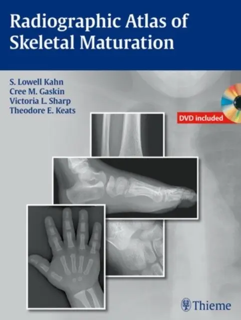
Radiographic Atlas of Skeletal Maturation
By S. Lowell Kahn
📘Hardback
$1153.99In stock
Code: 9781604065718
Radiographic Atlas of Skeletal Maturation
S. Lowell Kahn, Cree M. Gaskin, Victoria L. Sharp, Theodore E. Keats, 2012
Overview
This atlas provides a comprehensive reference for normal skeletal maturation, featuring nearly 2,300 high-quality images. It addresses the common clinical question of whether a skeletal image represents normal development or an alteration. The resource offers male and female reference images for every age and body part, aiding physicians in quickly identifying normal ranges. This is particularly valuable for interpreting pediatric skeletal radiographs in urgent situations.
Who it's for
- Physicians and radiologists evaluating pediatric skeletal development.
- Medical professionals needing to differentiate normal growth variations from injuries.
- Emergency room physicians on call for pediatric cases.
Key features
- Nearly 2,300 high-quality images illustrating normal skeletal development.
- Reference images available in both the text and an accompanying DVD.
- Multiple projections provided for each age, sex, and body part combination.
- Practical layout organized by gender and body part for quick access.
- Includes software with image enhancement tools and compatibility with DICOM format.
- Convenient growth charts included in both the book and DVD.